

Overview of Hip Anatomy
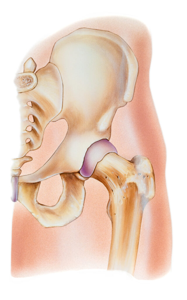
The hip is a ball-and-socket joint, which is one of the body’s largest and most important joints. The ball part of the joint is the femoral head, which is the upper end of the femur (thighbone). The socket is formed by the acetabulum, a part of the large pelvis bone. The hip joint’s anatomy is designed for stability and weight-bearing, with its articular cartilage, labrum, ligaments, muscles, and synovial fluid all contributing to its function and health.
Articular Cartilage and Labrum
The surfaces of the ball and the socket are covered by a slippery, robust tissue called articular cartilage. This cartilage creates a smooth, frictionless surface that helps the bones glide easily across each other during movement. The acetabulum is ringed by strong fibrocartilage called the labrum, which forms a gasket around the socket, deepening it and improving the stability of the joint.
Ligaments and Synovium
The hip joint is surrounded by bands of tissue called ligaments, which form a capsule that holds the joint together. The undersurface of the capsule is lined by a thin membrane called the synovium, which produces synovial fluid that lubricates the hip joint.
Muscles and Movement
Muscles around the hip joint are grouped based on their functions relative to the movements of the hip. These include flexors, extensors, adductors, abductors, internal rotators, and external rotators. The hip joint allows for movement in three major axes: flexion and extension, internal and external rotation, and abduction and adduction.
Stability and Blood Supply
Hip stability arises from several factors, including the shape of the acetabulum and the strength of the surrounding muscles and ligaments. The hip joint is also supplied by a network of blood vessels that provide essential nutrients to the joint structures.
Conditions and Treatments
Common hip problems include arthritis, bursitis, avascular necrosis, and femoroacetabular impingement (FAI). Treatments can range from conservative methods like physical therapy to surgical interventions such as total hip arthroplasty (THA) or periacetabular osteotomy (PAO) for joint preservation.
Hip Osteoarthritis
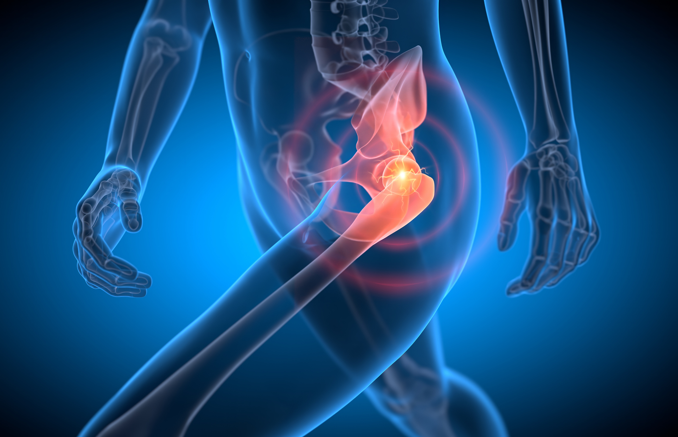
Manage Hip Osteoarthritis: Non-surgical treatments include weight management, therapy, medications, injections, devices, and heat therapy. Surgical options like arthroscopic debridement or total hip replacement may be needed if non-surgical methods fail. Post-surgery, rehabilitation with therapy and assistive devices aids recovery. Improve quality of life despite this chronic condition.
Avascular Necrosis of the Hip
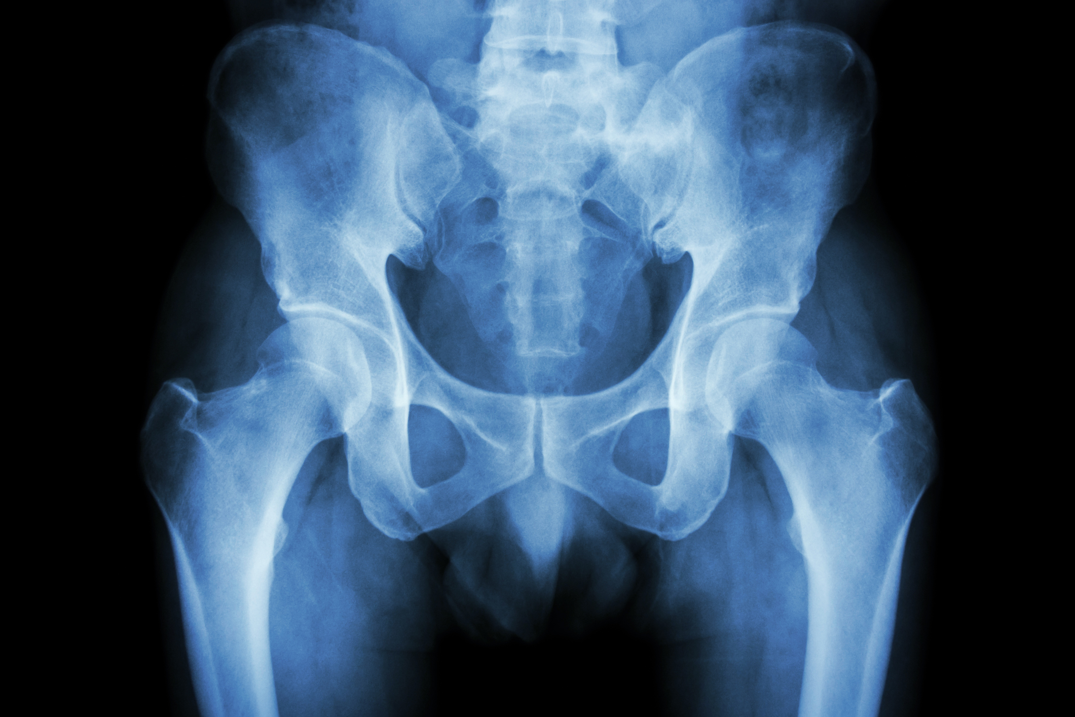
Avascular necrosis (AVN) of the hip, also known as osteonecrosis, is a condition characterized by the death of bone tissue due to a lack of blood supply. Although it can damage any bone, hip joints are the most frequently affected. An estimated 20,000 to 30,000 new instances of AVN [...]
Hip Labral Tears
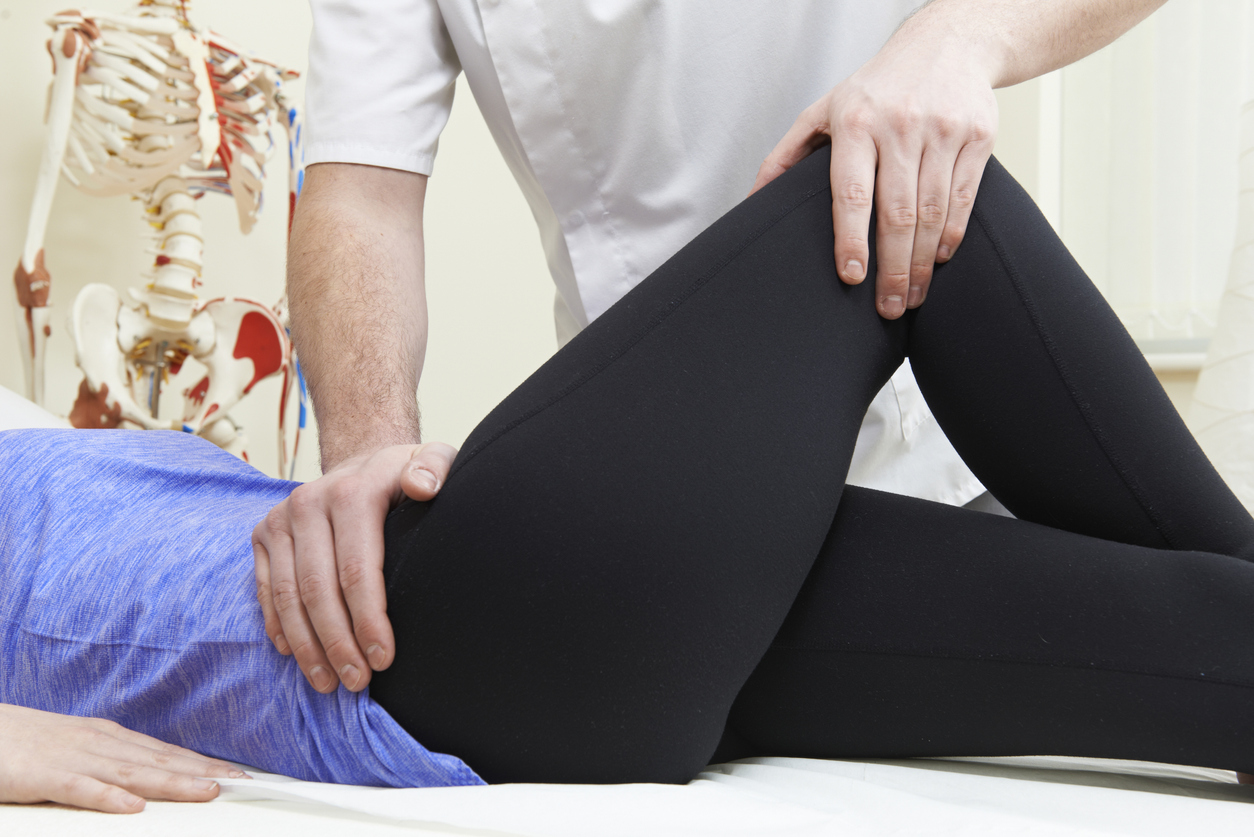
Hip Labral Tears: A Comprehensive Guide to Symptoms, Causes, and Treatments Understanding Hip Labral Tears Hip labral tears are a common yet complex condition affecting the hip joint, often leading to persistent pain and reduced mobility. The labrum, a ring of cartilage that encircles the hip socket, plays a [...]

Piriformis Syndrome
Diagnosing & Treating Piriformis Syndrome: Butt muscle compresses sciatic nerve, causing pain & numbness. Diagnosis via clinical exam & history review; MRI/CT to rule out other issues. Treat with rest, stretching, therapy, meds, or surgery. Prevent with exercise, good posture, avoiding prolonged sitting. Relief is possible with proper care.
Hamstring Tendon Tears
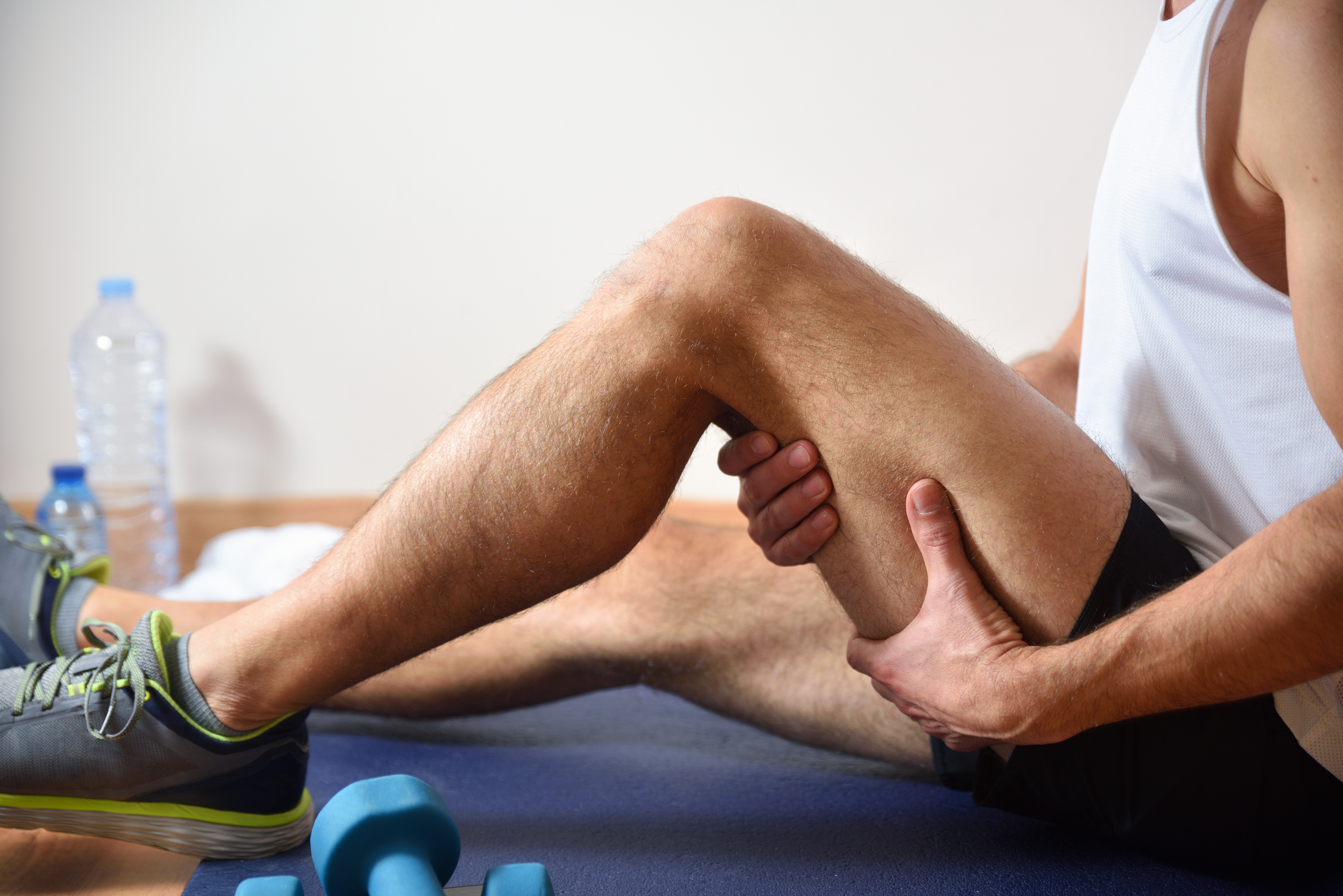
Understanding Hamstring Tendon Tears: Grades, Symptoms, and Treatment: Hamstring tendon tears range from mild strains to complete muscle tears, often affecting athletes. Classified into grades based on severity, treatment involves RICE therapy initially followed by gradual exercises. Symptoms include pain, stiffness, and swelling, with prevention strategies emphasizing stretching and warming up. Seeking medical advice is crucial for proper diagnosis and management, particularly for severe or recurring injuries. Learn more about managing hamstring injuries here.